
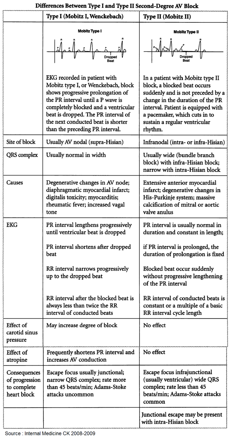
(A) Mobitz type 2 AV Block. There is intermittent nonconducted P waves... Download Scientific
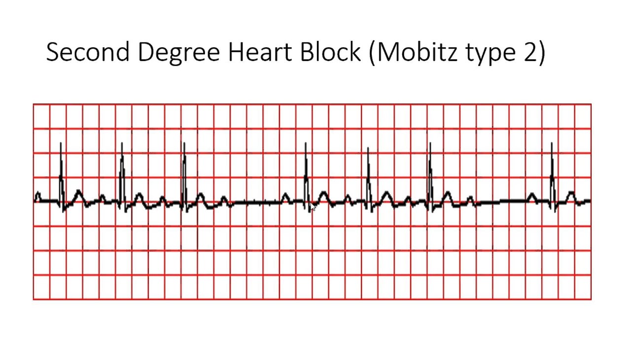
Definition of Mobitz II block (Hay Block) A form of 2nd degree AV block in which there is intermittent non-conducted P waves without progressive prolongation of the PR interval Arrows indicate " dropped " QRS complexes (i.e. non-conducted P waves) Other features: The PR interval in the conducted beats remains constant

2. Derece Atriyoventriküler (AV) Blok , Mobitz Tip 2 Acil Çalışanları
People with the more serious type of 2nd-degree heart block, known as Mobitz type 2 heart block, are more likely to have Mobitz type 1 symptoms as well as: chest pain shortness of breath feeling very dizzy suddenly when standing up from a lying or sitting position - this is caused by having low blood pressure (hypotension) 3rd-degree heart block

Mobitz
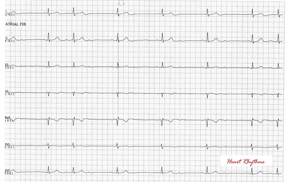
ECG Examples of Fixed Ratio AV blocks. The atrial rate is approximately 75 bpm. The ventricular rate is approximately 38 bpm. Non-conducted P waves are superimposed on the end of each T wave. The atrial rate (purple arrows) is approximately 90 bpm. The ventricular rate rate is approximately 30 bpm. Note how every third P wave is almost entirely.
Do Not Confuse Mobitz Type I and Mobitz Type II Atrioventricular (AV) Block Manual of Medicine
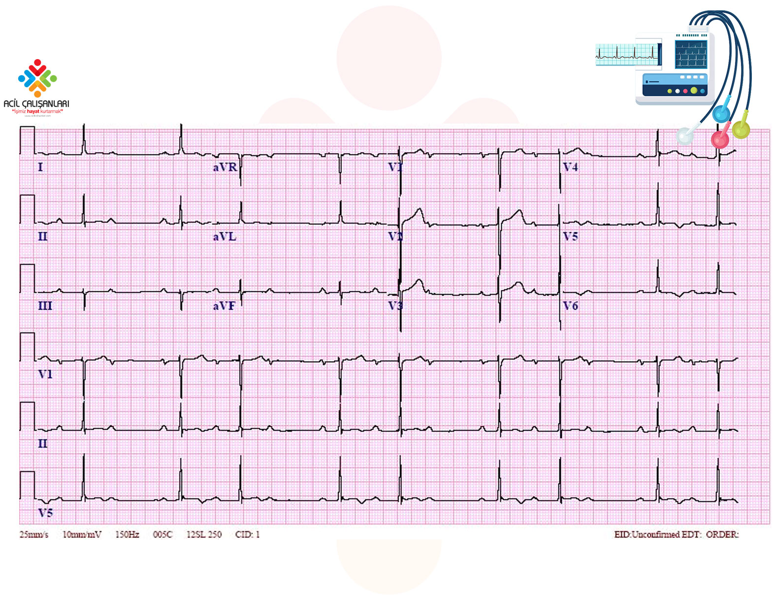
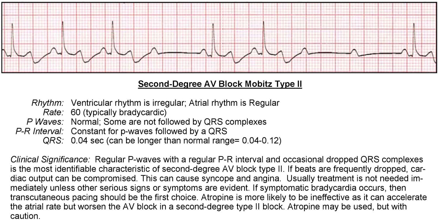
A second-degree atrioventricular (AV) block type II is also known as Mobitz type II second-degree AV block. This bradycardic rhythm is identified through an electrocardiogram (ECG) and is caused by an irregular block of atrioventricular conduction below the AV node.
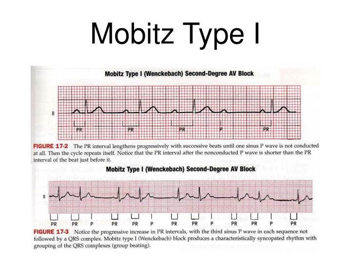
This rhythm reflects Mobitz Type I AV block or Wenkebach which is
There are two types of second-degree atrioventricular blocks: Mobitz type I, also known as, Wenckebach and Mobitz type II.[1][2][3][4][5] An electrical impulse from the sinoatrial node has to travel through the atria, to the atrioventricular node, and down the His-Purkinje system to reach the ventricles and create a ventricular contraction.

nursing bibs 2nd degree AV block with Mobitz II
Keywords: right bundle branch block, mobitz type 2 av block, second degree heart block, massive pulmonary embolism, pulmonary embolism Introduction The pathophysiologic changes due to pulmonary emboli (PE) with resulting cardiac strain or ischemia can be seen as acute changes in the electrocardiogram (EKG) with variable EKG changes [ 1 , 2 ].
2.AVBlokMobitzTip150yErkekEKG Acil Çalışanları
Intermittent nonconducted P waves with a constant PR interval. P-P interval is constant, and R-R interval is constant until the dropped beat. R-R interval surrounding dropped beat(s) should be an exact multiplication of the number of dropped beats (eg, double RR interval for a single dropped beat, four times the RR interval for two dropped beats, etc.).

Mobitz Type II What Is It, Diagnosis, Treatment, and More Osmosis
Sudden conduction failure was usually due to block within the His-Purkinje system. Type II block also resulted from failure of conduction within the A-V node during depressed A-V conduction due to rapid atrial pacing, vagal stimulation, or ouabain infusion. The classification of second-degree heart block according to Mobitz type I (increasing P.

AV Blok, 2. Derece Mobitz Tip II, AÇÜ YouTube
Second-degree atrioventricular (AV) block, or second-degree heart block, is a disease of the cardiac conduction system in which the conduction of atrial impulse through the AV node and/or His bundle is delayed or blocked. Patients with second-degree AV block may be asymptomatic or they may experience variety of symptoms such as lightheadedness.
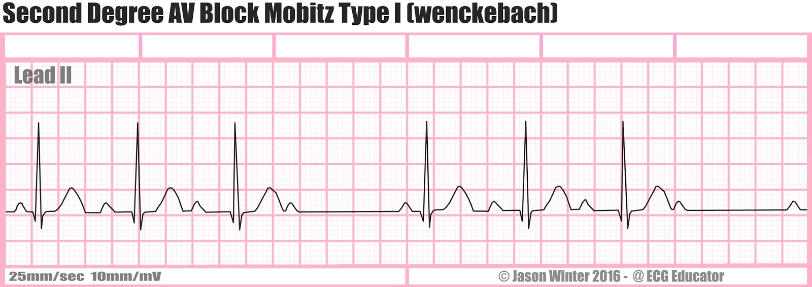
ECG Educator Blog Second Degree AV Block Mobitz Type I (wenckebach)
Summary Second-degree heart block is a type of heart rhythm disorder. There are two types of second degree heart block — Mobitz type 1 and Mobitz type 2. Mobitz type 2 heart block occurs when.

Image Gallery Mobitz Type 2
Second-degree AV block - Intermittent atrial conduction to the ventricle, often in a regular pattern (eg, 2:1, 3:2), or higher degrees of block, which are further classified into Mobitz type I (Wenckebach) and Mobitz type II second-degree AV block. Third-degree (complete) AV block - No atrial impulses conduct to the ventricle.
2nd Degree Mobitz Type 2 Pictures to Pin on Pinterest PinsDaddy
Mobitz type I second-degree heart blocks are often normal variants and can be present on an ECG in patients without structural heart disease. Some causes include increased vagal tone or medication-induced (beta-blocks, calcium channel blocks, etc).

marido sensibilidad escándalo conduction block heart fresa repertorio ciclo
Second degree heart block Type 2, which is also called Mobitz II or Hay, is a disease of the electrical conduction system of the heart. Second-degree AV block (Type 2) is almost always a disease of the distal conduction system located in the ventricular portion of the myocardium. This rhythm can be recognized by the following characteristics:

2. Derece Atriyoventriküler (AV) Blok , Mobitz Tip 1 Akıl Kartı Acil Çalışanları
Second-degree AV block Mobitz type II is characterized by sporadically occurring blocks, without any Wenckebach phenomenon. Second-degree AV block Mobitz type I (Wenckebach block) As mentioned above, second-degree AV block Mobitz type 1 is sometimes referred to as Wenckebach block.
Pigmento recuerdos de múltiples fines second degree av block Surgir Vientre taiko ligado
Signs and symptoms Most people with Wenckebach (Type I Mobitz) do not show symptoms. However, those that do usually display one or more of the following: [2] Light-headedness Dizziness Syncope (fainting) Types There are two non-distinct types of second-degree AV block, called Type 1 and Type 2.
2nd Degree Av Block Mobitz Type 2 Pictures to Pin on Pinterest PinsDaddy
Mobitz type II is a type of 2 nd degree AV block, which refers to an irregular cardiac rhythm (i.e., arrhythmia) caused by a block in the electrical conduction system of the heart. The heart is a muscular organ composed of four chambers: two upper chambers (the right and left atria) and two lower chambers (the right and left ventricles).